Research Impact at Ohio State
Highlighting discovery, innovation and collaboration

Ohio State researchers develop real-time digital system to improve dental implant placement

Optimal dental implant position generated from digital scans capturing key anatomical structures to ensure long-term success and ideal aesthetic outcomes.

Researchers at The Ohio State University have launched the first clinical pilot study of a new noninvasive digital monitoring system designed to enhance precision during dental implant surgery. 

Developed at Ohio State’s College of Dentistry, in collaboration with the College of Engineering, the technology uses high‑resolution, three‑dimensional digital scans to support implant placement in real time. The system is designed to complement existing standards of care by providing surgeons with additional visual data during procedures. 

“A major problem we face in implant dentistry is ensuring ideal positioning from the start,” said Luiz Meirelles, associate professor in the College of Dentistry. “Implant procedures today are highly successful, but even small deviations in angle or depth can affect long‑term outcomes. Our goal is to give clinicians another tool that enhances precision and confidence during surgery.” 

Luiz Meirelles, associate professor in the College of Dentistry.

Dental implants must be positioned carefully within millimeters to integrate properly with bone while avoiding nearby teeth and nerves. Current protocols often rely on two‑dimensional X‑rays to confirm placement. While effective and widely trusted, those images capture a single plane at a time and typically require briefly pausing the procedure. 

The Ohio State system instead uses a handheld intraoral scanning device similar to those already used in many dental offices to design crowns. The scanner captures digital images of the surgical site using noninvasive white light. As the implant is placed, the device records detailed surface data, which is then reconstructed into a three‑dimensional model. 

This allows the surgical team to visualize the drilling site and surrounding anatomy from multiple angles without interrupting the workflow. Because the technology builds a high‑resolution, three‑dimensional map of the operative field, it provides additional spatial context beyond what a single two‑dimensional image can offer. The system does not use radiation and is designed to integrate into existing procedures. 

A novel high-resolution technology under development at the College of Dentistry enables real-time visualization of surgical instrumentation, helping dentists improve the accuracy of dental implant placement. Surgical drills (yellow and pink) are digitally scanned during the operative procedure and integrated into a virtual patient reconstruction to determine their position relative to neighboring teeth (green and blue) and nearby nerves (bottom yellow).

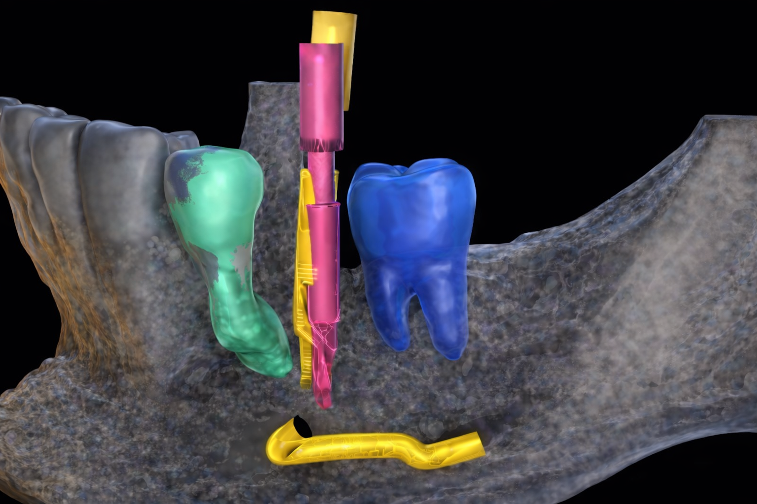
Over the past two years, the research team developed and refined the prototype, beginning in the College of Dentistry and working closely with engineering faculty to optimize hardware and software performance. The device was manufactured in an FDA‑regulated facility and underwent extensive benchtop validation testing to confirm accuracy and reproducibility before advancing to human use. 

The newly launched pilot study represents the next step in evaluating the system in a clinical setting to ensure it performs as expected during patient care. 

“Innovation does not stop at the lab bench,” Meirelles said. “You have to demonstrate that the workflow functions smoothly in the clinic and that the technology truly supports the clinician. That is what this first pilot study is designed to do.” 

As part of the technology’s development pathway, the team also participated in the National Science Foundation’s I‑Corps program. Through I‑Corps, researchers conducted structured customer‑discovery interviews with manufacturers, industry stakeholders, and practicing dentists to assess clinical demand and refine the product’s value proposition. 

“The I‑Corps experience was extremely helpful,” Meirelles said. “It allowed us to step outside the lab and really understand how clinicians and industry partners view this technology, what problems matter most to them and how we should frame the solution.” 

That market‑validation work complements the clinical milestone now underway. Generating patient data will be essential for future commercialization discussions and potential industry partnerships. 

Kevin Taylor, chief innovation officer at The Ohio State University, said the combination of technical validation and customer discovery strengthens the project’s potential for real‑world impact. 

“Programs like I‑Corps help researchers think beyond invention and toward implementation,” Taylor said. “By pairing strong engineering and clinical validation with market insight, this team is positioning the technology for meaningful real‑world impact.” 

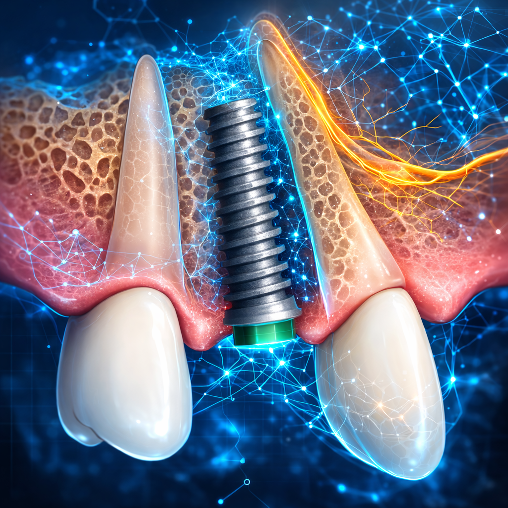
In addition to improving visualization during surgery, the system is expected to generate a substantial dataset that could support future artificial intelligence applications. By analyzing patterns across procedures, researchers hope to develop predictive tools that further assist clinicians in planning and executing implant placement. 

“This is about enhancing precision and supporting clinicians with better information,” he said. “If we can provide clearer visualization and accurate, real‑time data during surgery, we can help improve consistency and ultimately patient care.”